Dentoalveolar

Trevor Oliverson, MD, DDS
Oral and Maxillofacial Surgery Resident
Mayo Clinic
Rochester, Minnesota, United States

An Qi Wu, MD, DDS
Oral and Maxillofacial Surgery Resident
Mayo Clinic
rochester, Minnesota, United States
James Van Ess, MD, DDS
Oral and Maxillofacial Surgeon
Mayo Clinic
Rochester, Minnesota, United States
.png) Figure 2. Case 1 – Intraoperative Photographs. A) Site #9 following osteotomy preparation with B) 3D-printed donor tooth replica used for surgical planning. C) Extraction site of the partially erupted tooth #8a and D) Donor tooth fixated into site #9 with a composite splint.
Figure 2. Case 1 – Intraoperative Photographs. A) Site #9 following osteotomy preparation with B) 3D-printed donor tooth replica used for surgical planning. C) Extraction site of the partially erupted tooth #8a and D) Donor tooth fixated into site #9 with a composite splint.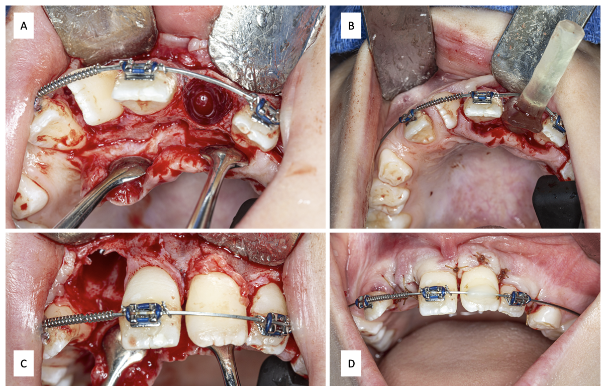 Figure 3. Case 1 – Postoperative Presentation. A) Panoramic radiograph at 6 weeks postoperatively, demonstrating appropriate positioning of tooth #8a in site #9. B) Clinical image following Phase I orthodontics at 12 months, showing favorable movement of the anterior dentition, appropriate space closure, and correction of asymmetry.
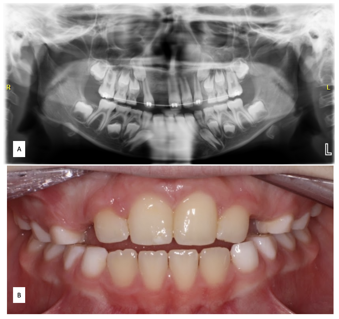